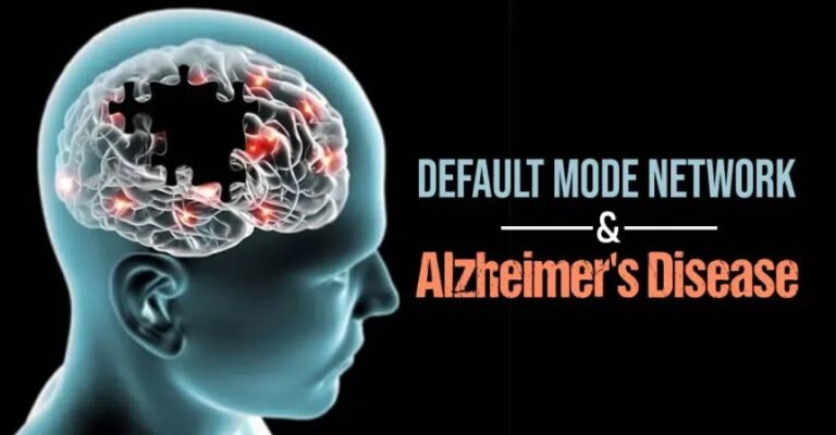
The default mode network (DMN) is strongly associated with a wide range of conditions such as Alzheimer’s disease (AD). Both DMN and AD involve two main brain regions, namely the posterior cingulate cortex and hippocampal formation.
What Is The Default Mode Network?
The default mode network (DMN) refers to an arrangement of connected brain areas that display an increased activity when a person is in a state of wakeful rest and not focused on the outside world. The default mode network plays a crucial role in regulating the self-referential introspective state [mfn] Mak LE, Minuzzi L, MacQueen G, Hall G, Kennedy SH, Milev R. The Default Mode Network in Healthy Individuals: A Systematic Review and Meta-Analysis. Brain Connect. 2017 Feb;7(1):25-33. doi: 10.1089/brain.2016.0438. Epub 2017 Jan 9. PMID: 27917679. [/mfn] and is associated with a range of brain regions.
According to a 2018 study [mfn] Davey, C. G., & Harrison, B. J. (2018). The brain’s center of gravity: how the default mode network helps us to understand the self. World psychiatry : official journal of the World Psychiatric Association (WPA), 17(3), 278–279. https://doi.org/10.1002/wps.20553 [/mfn] , “The DMN is composed primarily of medial prefrontal cortex (MPFC) and posterior cingulate cortex (PCC), both situated along the brain’s midline, together with inferior parietal and medial temporal regions.” DMN activity increases during perspective-taking of the desires, beliefs, and intentions of others, in remembering the past, and in planning the future.
These functions genetically involve the self as a reference point. The self-referential properties of these functions suggest that the default mode network may contribute to adaptive behavior by allowing scenarios to be constructed, replayed, and explored in the mind, both to ponder past events and to derive expectations about the future. Failure to overcome DMN activity may result in interference from internal mentation [mfn] Andrews-Hanna, J. R. (2011). The brain’s default network and its adaptive role in internal mentation. The Neuroscientist, 18(3), 251-270. https://doi.org/10.1177/1073858411403316 [/mfn] (i.e. the introspective & adaptive mental activities) or emotional processing, which we deliberately and spontaneously engage in regularly.
Read More About Default Mode Network Here
Alzheimer’s Disease And Default Mode Network
Alzheimer’s disease (AD) is a progressive and permanent brain disorder [mfn] Schachter, A. S., & Davis, K. L. (2000). Alzheimer’s disease. Dialogues in clinical neuroscience, 2(2), 91–100. https://doi.org/10.31887/DCNS.2000.2.2/asschachter [/mfn] that impairs thinking skills, memory and other cognitive abilities that affect a person’s ability to function in daily life. Symptoms usually tend to appear during the patient’s mid-60s. A research paper [mfn] Kumar A, Sidhu J, Goyal A, et al. Alzheimer Disease. [Updated 2020 Nov 18]. In: StatPearls [Internet]. Treasure Island (FL): StatPearls Publishing; 2021 Jan-. Available from: https://www.ncbi.nlm.nih.gov/books/NBK499922/ [/mfn] explains that AD is the most common reason for cognitive decline. “It is a neurodegenerative disorder that usually affects people over the age of 65 with the involvement of language, memory, comprehension, attention, judgment and reasoning,” add the researchers. Research [mfn] Mevel, K., Chételat, G., Eustache, F., & Desgranges, B. (2011). The default mode network in healthy aging and Alzheimer’s disease. International Journal of Alzheimer’s Disease, 2011, 1-9. https://doi.org/10.4061/2011/535816 [/mfn] reveals that healthy aging and Alzheimer’s [mfn] Wu, X., Li, R., Fleisher, A. S., Reiman, E. M., Guan, X., Zhang, Y., Chen, K., & Yao, L. (2011). Altered default mode network connectivity in Alzheimer’s disease-A resting functional MRI and bayesian network study. Human Brain Mapping, 32(11), 1868-1881. https://doi.org/10.1002/hbm.21153 [/mfn] are associated with in the DMN, which is often described in people at risk for AD.
Studies [mfn] Beason-Held L. L. (2011). Dementia and the default mode. Current Alzheimer research, 8(4), 361–365. https://doi.org/10.2174/156720511795745294 [/mfn] also show that people suffering from age-related cognitive impairments, such as Alzheimer’s, tend to have changes associated with the default network, specifically in the hippocampal and posterior cingulate/precuneus regions. According to a 2004 study [mfn] Greicius, M. D., Srivastava, G., Reiss, A. L., & Menon, V. (2004). Default-mode network activity distinguishes Alzheimer’s disease from healthy aging: evidence from functional MRI. Proceedings of the National Academy of Sciences of the United States of America, 101(13), 4637–4642. https://doi.org/10.1073/pnas.0308627101 [/mfn] , the hippocampus is involved in the default mode network, and DMN activity is “deficient in AD compared to healthy elderly controls, and a metric of network activity shows promise as a clinical marker of AD.” A 2018 study [mfn] Grieder M, Wang DJJ, Dierks T, Wahlund LO, Jann K. Default Mode Network Complexity and Cognitive Decline in Mild Alzheimer’s Disease. Front Neurosci. 2018 Oct 23;12:770. doi: 10.3389/fnins.2018.00770. PMID: 30405347; PMCID: PMC6206840. [/mfn] further found that cognitive decline linked with AD is reflected by reduced complexity in DMN node signals and this can cause “disrupted DMN functional connectivity.”
Read More About Alzheimer’s Disease Here
How DMN And Alzheimer’s Are Connected?
The human resting-state is defined by spatially consistent brain activity at a low temporal frequency. The default mode network (DMN) is one of the so-called resting-state networks. It is associated with cognitive processes that are directed toward the self, such as introspection and autobiographical memory. The DMN’s integrity appears to be crucial for mental health.
Patients with Alzheimer’s disease (AD) have displayed disorders of functional connectivity within the brain regions of the DMN. However, in the early stages of Alzheimer’s disease, physiological alterations are sometimes baffling, despite manifested cognitive impairment. While functional connectivity estimates the signal correlation between brain areas, multi-scale entropy (MSE) measures the complexity of the blood-oxygen-level dependent signal within an area and thus might show local changes before connectivity is affected.
A study states that when it comes to AD, default network activity changes are hand in hand with those found using FDG-PET measure of resting-state brain metabolism. These highlights the major involvement of the PCC/precuneus region for positron emission tomography (PET) studies. For instance, the functional connectivity between the PCC and the hippocampus seems to be impaired in AD, presumably as a result of early hippocampal structural alterations. This so-called disconnection hypothesis has gained strong support from previous works combining structural MRI and PET.
Thus, hippocampal atrophy seems to provoke PCC functional perturbation, as well as episodic memory impairment, through interruption of the cingulum bundle. Resting-state fMRI studies have displayed alterations of the temporal synchrony of PCC and hippocampus activity in patients with aMCI, compared to healthy controls which are consistent with this hypothesis. Decreases in functional connectivity or deactivation disturbances have also been recorded within the PCC of aMCI patients and have been interpreted as the outcome of local atrophy.
Alteration of DMN activity in AD is not only limited to the PCC and hippocampal region as connectivity disturbance between these structures and other brain areas have also been observed. Such disruptions also seem to spread within the cortex as the disease progresses, that is, respectively, from aMCI to AD and from mild to severe AD.
Consistently, aMCI deactivations were less marked in the precuneus and medial frontal regions, while in AD patients, deactivations were restricted to medial frontal areas. As proposed in normal aging, these findings are believed to reveal a difficulty to switch from a resting-state to a task-related mode of brain function, mainly due to a failure of DMN brain regions to show rapid and effective coordination in their activity. Improvements in DMN activity or connectivity have also been reported in patients with aMCI or AD as compared to healthy aged controls.
Thus, aMCI patients were found to be characterized by:
- Progress in DMN activity located within the PCC/precuneus, (pre)frontal, lateral parietal, and middle temporal cortices
- Developments in functional connectivity between the right parietal cortex and left insula
Improvements in AD patients were found in regards to:
- DMN activity within the PCC/precuneus, frontal, occipital, parietal, and (pre)frontal cortices
- DMN connectivity between the left hippocampus and prefrontal dorsolateral cortex or between PCC and left frontoparietal cortices.
Altogether, these results point to the presence of possible compensatory processes appearing in the early stages of the disease and found in several DMN areas. It is worth mentioning that cognitive reserve was found to modulate the effect of pathology on brain function in general and on DMN activity in particular.
Effect Of Memantine On DMN In Alzheimer’s
According to a 2011 research [mfn] Lorenzi M, Beltramello A, Mercuri NB, Canu E, Zoccatelli G, Pizzini FB, Alessandrini F, Cotelli M, Rosini S, Costardi D, Caltagirone C, Frisoni GB. Effect of memantine on resting state default mode network activity in Alzheimer’s disease. Drugs Aging. 2011 Mar 1;28(3):205-17. doi: 10.2165/11586440-000000000-00000. PMID: 21250762. [/mfn] , memantine is a recommended, symptomatic treatment approach for moderate to severe Alzheimer’s disease. It reduces the excitotoxic effects of hyperactive glutamatergic transmission. However, the specific mechanism of the impact of memantine [mfn] McShane, R., Westby, M. J., Roberts, E., Minakaran, N., Schneider, L., Farrimond, L. E., Maayan, N., Ware, J., & Debarros, J. (2019). Memantine for dementia. The Cochrane database of systematic reviews, 3(3), CD003154. https://doi.org/10.1002/14651858.CD003154.pub6 [/mfn] in Alzheimer’s disease patients is inadequately understood.
It is essential to note that the default mode network (DMN), a brain system that plays a key role in attention, is hypoactive in Alzheimer’s disease and is under glutamatergic control. The study suggests “a positive effect of memantine treatment in patients with moderate to severe Alzheimer’s disease, resulting in an increased resting DMN activity in the precuneus region over 6 months.” Currently, memantine is the only approved [mfn] Robinson DM, Keating GM. Memantine: a review of its use in Alzheimer’s disease. Drugs. 2006;66(11):1515-34. doi: 10.2165/00003495-200666110-00015. PMID: 16906789. [/mfn] pharmacotherapeutic agent for treating moderate to severe AD.


Leave a Reply